| |
The entirety of the STG has only 30 cell
bodies, and 14 of these cells are located in the pyloric circuit.
Each of these cells is then categorized into six broad categories:
Anterior burster (AB), Pyloric dilator (PD), Pyloric (PY),
Inferior cardiac (IC), Ventricular dilator (VD), and Lateral
pyloric (LP). The AB
is the “pacemaker”– it is the only tonically bursting cell
in the circuit. That is, it will continue producing action
potentials without any sensory or synaptic input. In the event
that all the neurons are oscillating in the circuit, it is the AB
cell that has the fastest oscillation, pushing the other neurons
to its frequency. The PD cell controls the cardiopyloric valve,
and is a pacemaker by virtue of its electrical synapse with the AB
cell. The VD and IC cells control the ventral stomach grooves.
Finally the 8 PY cells and the LP cell controls pyloric filter
movements.
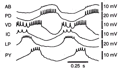
In the following explanation of the mechanism
of the pyloric circuit, refer to the circuit diagram below:
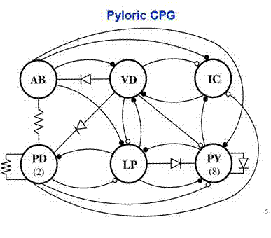
Initially, the AB cell, which is phasically
bursting, forces the electrically coupled PD cells into synchrony.
Note that the PD cells are tonically active, and only
display the bursting pattern due to the electric coupling with the
AB cell. These three cells inhibit the 8 PY cells, causing them to
fire out of sync (these cells can only fire when the AB and PD
cells are not inhibiting them, that is to say when they are
inactive). The VD, LP,
and IC cells are rebound spikers, so they spike as a result of a
strong inhibition. Each of these cells then has inhibiting
interactions with other cells in the circuit, causing the specific
voltage patterns shown below.
|